Field Notes · October 11, 2025 · 6 min · By Esperanza Whitford
The microscopic mapping that makes Mohs precise
How the surgeon turns your tumor into a map and reads every margin.
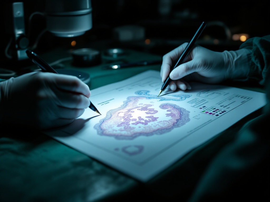
What sets Mohs micrographic surgery apart is not just that it checks margins, but how meticulously it maps and reads them, a process worth understanding because it explains the exceptional cure rate.
After removing a thin layer of tissue, the surgeon divides and color-codes it, drawing a precise map that records the exact orientation of every edge relative to the patient. The tissue is then frozen, sectioned, and examined under the microscope so that essentially the entire margin, both the deep surface and all the edges, is inspected, rather than the small sampling a standard pathology cut provides. If cancer appears at a specific mapped point, the surgeon knows precisely where on the patient to remove more, and only there.
This mapping is the engine of Mohs. Standard excision examines a fraction of the margin and reports days later; Mohs examines the whole margin on-site and acts on it immediately and locally. The result is both the near-complete certainty that no tumor is left and the conservation of healthy tissue, since removal is guided to exactly where cancer is. The long appointment reflects this careful processing and reading, the thoroughness that delivers the result, and the reason for the cure rate so often quoted.
Related reading: Aggressive skin cancer subtypes and why Mohs matters.